Day 1 :
Keynote Forum
Wassil Nowicky
Nowicky Pharma/ Ukrainian Anti-Cancer Institute, Austria
Keynote: The antiangiogenic properties of NSC-631570 (Ukrain) on breast cancer

Biography:
Dr. Wassil Nowicky — Dipl. Ing., Dr. techn., DDDr. h. c., Director of “Nowicky Pharma” and President of the Ukrainian Anti-Cancer Institute (Vienna, Austria). Has finished his study at the Radiotechnical Faculty of the Technical University of Lviv (Ukraine) with the end of 1955 with graduation to “Diplomingeniueur” in 1960 which title was nostrificated in Austria in 1975. Dr. Wassil Nowicky became the very first scientist in the development of the anticancer protonic therapy and is the inventor of the preparation against cancer with a selective effect on basis of celandine alkaloids “NSC-631570”. He used the factor that cancer cells are more negative charged than normal cells and invented the Celandine alkaloid with a positive charge thanks to which it accumulates in cancer cells very fast. Author of over 300 scientific articles dedicated to cancer research. Dr. Wassil Nowicky is a real member of the New York Academy of Sciences, member of the European Union for applied immunology and of the American Association for scientific progress, honorary doctor of the Janka Kupala University in Hrodno, doctor “honoris causa” of the Open international university on complex medicine in Colombo, honorary member of the Austrian Society of a name od Albert Schweizer. He has received the award for merits of National guild of pharmasists of America. the award of Austrian Society of sanitary, hygiene and public health services and others.
Abstract:
One of important features of anti-cancer drug NSCâ€631570 is the inhibition of the formation of the new blood vessels supplying a tumor. Due to these antiangiogenic properties NSC-631570 administered before surgery brings about better demarcation of the tumor from surrounding tissue and the tumor encapsulation. This alleviates the surgical removal of tumors what has been confirmed in breast cancer studies. In tests in vitro, NSC-631570 inhibited in a dose dependent manner the proliferation of human endothelial cells without exerting cytotoxic effect. The angiogenesis inhibition was observed on the capillary formation model. This inhibition of the neoangiogenesis prevents the metastasis formation as well.
The researchers from Emory University (Atlanta, Georgia, USA) and Kennesaw State University (Kennesaw, Georgia, USA) concluded: “The anticancer drug Ukrain exerts its cytotoxic effects on both mouse and human breast cancer cell lines in a dose and time dependent manner. Weeks following Ukrain treatment, cells maintained a reduced capacity to proliferate. Our data suggest that Ukrain could be effective as an anticancer drug for breast cancer due to its short term and long term inhibitory effects on tumor cell viability and proliferation.” This work was supported by RO1 CAâ€138993 and the NSF Award # 0450303 Subaward # 1â€66â€606â€63.
- Sessions: Screening, Detecting and Diagnosing Breast Cancer | Breast Cancer Therpy , Prevention and Medicine
Location: Odenwald
Session Introduction
Manuela Lacerda
IPATIMUP, Portugal
Title: Concerning the changing of diagnostic approach, guidelines, role and importance on decision of the different medical specialties involved ( in the last 30 years) in the setting of a screening and treatment reference hospital
Time : 11:55-12:25
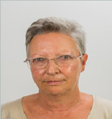
Biography:
Maria Manuela Pinto de Lacerda (Manuela Lacerda -name used in scientific publications) is specialist in surgical pathology. She started working at Instituto Português do Oncologia Francisco Gentil (IPOFG), Coimbra, Portugal in 1978. From 1986 to 2009 Director of the Laboratory Department of IPOFG, Coimbra, Portugal. From 2009 to 2011 Director of the Service of Surgical Pathology of IPOFG, Coimbra, Portugal. From 1989 to 1995 was part of the Technical Team of the Regional Cancer Registry - Central Zone (R.O.R.Centro), Portugal.from 1998 to 2001 Clinical Director of IPOFG, Coimbra, Portugal. From 1994 to 2011, was part the European Working Group on Breast Screening Pathology, which have developed "European Guidelines for Quality assurance in breast cancer screening and diagnosis" - European Commission. Since 2012 Consultant at Instituto de Patologia e Imunologia Molecular da Universidade do Porto (IPATIMUP), Porto, Portugal. She did her PhD in 2005 from Faculty of Medicine, Porto, Portugal. She is co-author of 6 papers in national journals with scientific arbitration and 20 articles in journals of international circulation with scientific arbitration.
Abstract:
In 1978 I started working, as surgical pathologist, in the cancer hospital- Instituto Português de Oncologia Francisco Gentil (IPOFG) - Coimbra, Portugal.
At that time the patients that came to the hospital had large breast tumors and the diagnosis was made by the “triple approach”: The members of the multidisciplinary team were a surgeon, a radio-therapist and a medical oncologist. The pathology report was descriptive.
Frozen section or biopsy of the lesion was mandatory when the first therapeutic approach was surgery or when there was no concordance in the triple approach.
In 1987 the papers and the book published by David L Page and William D. Dupont, became a landmark in the histoclinical approach of breast lesions. The reproducibility of the prognostic index of Nottingham proposed by Elston and Ellis was an important method for the follow-up of breast cancer.
Immunohistochemistry became routine either for differential diagnostic or for therapeutic guidance.
In 1990 a breast cancer screening program in the central zone of Portugal was implemented. It was one of the European pilot projects approved by the European Commission. This program covers a female population (age groups 45-70) of 320,225 women (mammography every two years).
In 1994 I was designated, by the management of the program, to integrate the European Working Group on Breast Screening Pathology that elaborated "European Guidelines for Quality assurance in breast cancer screening and diagnosis - Quality Assurance guidelines for pathology" - European Commission.
The Cancer Registry -Central Zone (R.O.R. –Zona Centro) data show the influence of breast cancer screening on the incidence and mortality of breast cancer.
Alicia Gonzalez Gonzalez
University of Cantabria, Spain
Title: Melatonin enhances chemosensitivity of human endothelial and breast cancer cells by regulating genes involved in angiogenesis
Time : 12:25-12:55

Biography:
Alicia Gonzalez Gonzalez is a PhD student from Cantabria University School of Medicine. Currently, she is involved in Breast Cancer research and Melatonin, specifically in the sensitizing effects of melatonin to chemotherapy and radiotherapy for its antiangiogenic and antiadipogenic actions. She has published three papers and she has presented her work in five international conferences.
Abstract:
Endothelial cells represent one of the critical cellular elements in tumor microenvironment playing a crucial role in the growth and progression of cancer through controlling angiogenesis. Enhancing the chemosensitivity of cancer cells is one of the most important goals in clinical chemotherapy. Melatoanin exerts oncostatic activity in breast cancer through antiangiogenic actions. Vascular endothelial growth factor (VEGF) produced from tumor cells is essential for the expansion of breast cancer. The angiopoietin/Tie-2 family play an important role in regulating vessel stability. Several studies emphasize the complementary and coordinated roles of angiopoietin-2 and VEGF during angiogenesis. Thus, the aim of the present study was to investigate whether melatonin sensitizes endothelial cells to chemotherapy by regulating angiogenesis. To accomplish this we used human umbilical vein endothelial cells (HUVEC) or cocultures of HUVEC cells with MCF-7 cells. Cell proliferation was measured by the MTT method. We selected different genes which were modulated by melatonin from an array of genes involved with the process of angiogenesis. Their expression was analyzed by RT-PCR. The migration of HUVECs was measured by the Wound Healing Assay and tubulogenesis studies were performed in a tubulogenesis multiplate system in vitro. Only the presence of malignant epithelial cells in the cocultures altered proliferation, and stimulated ANG-1, ANG-2 and VEGF expression and decreased Tie2 expression in endothelial cells. Melatonin 1 mM added to the coculture counteracted this effect and reduced Ang-1, Ang-2 and VEGF expression and increased Tie-2 expression. In addition, vinorrelbine and docetaxel decreased cell proliferation and melatonin led to a significantly greater decrease. Furthermore, it is important to point out that chemotherapy increases the expression of genes involved in angiogenesis such as VEGF, ANG1, ANG2, FGFR3, NOS3 or MMP14, and melatonin pretreatment 1 mM led to a significantly decrease. Furthermore, the migration of endothelial cells and the tube formation were reduced with the chemotherapy and melatonin pretreatment resulted in a significantly higher reduction. All these results suggest that melatonin could exert a cooperative enhancement of chemosensitivity associated with the modulation of angiogenesis. Therefore, melatonin could represent a new and promising therapeutic approach to the treatment of breast cancer.
Javier Menéndez Menéndez
University of Cantabria, Spian
Title: Effects of melatonin on chemotherapy treatments in gene expression and proteomics of human breast cancer

Biography:
Javier Menéndez Menéndez is a young researcher in Molecular Biology and Biomedicine who is completing his PhD from Cantabria University. He is investigating about breast cancer and the sensitizing effects of melatonin in chemotherapy and radiotherapy since 2014. He has published five papers in reputed journals as well as a book chapter. Also, he has presented his works in two national congresses.
Abstract:
Melatonin is the main secretory product of the pineal gland. It is an oncostatic agent that reduces the growth and development of various types of tumors, particularly those whose growth is dependent on estrogen. Data from nearly 500 articles published in the last 20 years, call attention to the hypothesis that melatonin interacts with estrogen signaling pathways at three different levels: 1) Acting as a selective estrogen receptor modulator (SERM), obstructing the activation of estradiol receptors, and consequently, down regulating the expression of proto- oncogenes, growth factors and genes involved in process like proliferation, invasiveness and metastasis. 2) Acting as a selective modulator of the enzymes involved in the synthesis of steroid hormones (SEEM), inhibiting the expression of some enzymes (aromatase and sulfatase estrogen) and stimulating others (estrogen sulfotransferase). 3) Interfering with the hypothalamus-pituitary-reproductive axis in such way that the synthesis of estrogens is decreased. Correspondingly to the properties mentioned, melatonin might be a very good adjuvant for chemotherapy treatments currently used in cancer. However, little is known about the consequences that melatonin might have over the molecular changes induced by chemotherapy agents at the tumor cell level. In this work, as an experimental approach, we cotreated an estrogen receptor positive cell line derived from a mammary adenocarcinoma (MCF-7 cells) with chemotherapy agents and the pineal hormone, in order to investigate whether melatonin can modulate the gene expression and the proteomic changes induced by the chemotherapy agents.
Cankaya Baris
Marmara University Pendik Training Hospital, Turkey
Title: Blocks for relieving pain associated with breast surgery

Biography:
I am anesthesiologist with interest in perioperative medicine and patient safety. I am responsible for blue code management in my hospital. I have certifications for adult, newborn, pediatric resuscitation from Europen Resuscitation Council.
Abstract:
Regional and neuraxial anesthesia for pain management after breast surgery is gaining necessity. Data show improved postoperative pain control and patient satisfaction scores. Acute postoperative pain is a risk factor for chronic pain in women after breast surgery. Chronic postoperative pain develops in nearly half of patients undergoing breast surgery. Nerve blockade improves postoperative analgesia with decreased volatile anesthetic use and decreases hospital length of stay. most commonly performed procedures are thoracic epidural catheters
and paravertebral blocks , also ultrasound guided interfascial
plane blocks that target Pectoral nerve (Pecs) are Pecs I (between the pectoralis majör) Pecs II ( between the pectoralis minor and serratus anterior
muscles). The local anesthetic blocks the medial and lateral pectoral nerves, anterior divisions of the thoracic intercostal nerves from T2 to T6, long thoracic nerve, and
thoracodorsal nerves thus providing analgesia. PECs blocks have
shown efficacient for analgesia after breast surgery. PECs easy to administer and associated with a lower incidence of complications, especially with the use of ultrasonography. Pecs block has been performed as postoperative pain management; not for a primary anesthesia. Anesthesiologists are increasingly prefering Pecs over Thoracal Paravertebral blocks and thoracic epidural catheters. PECs have lower risk of intravascular injection.
Gul Cankaya
Marmara University Pendik Training Hospital, Istanbul/ Turkey
Title: The breast-q in perioperative period for breast conserving surgery: how patient reported outcome measures contribute to health related quality of life

Biography:
Gul CANKAYA has been working as a surgery nurse for 4 yrs. She is working on management for breast cancer patients during perioperative period. She has completed international breast care nursing training program. She has interest in palliative care and has trainer certification.
Abstract:
Cankaya Gul, Surgery Nurse , National Health Service Marmara University Pendik Training Hospital Istanbul/ TURKEY
Advances in breast cancer management have reduced breast mortality rates.
The BREAST-Q is a patient reported outcome measurement (PROM) owned by
Memorial Sloan-Kettering Cancer Center and the University of British Columbia.
This questionnaire consists of six key themes of patient satisfaction and health-related quality of life in breast surgery:
1. Satisfaction with breasts
2. Satisfaction with overall outcome
3. Psychosocial well-being
4. Sexual well-being
5. Physical well-being and
6. Satisfaction with care
Breast augmentation is the most common cosmetic surgery procedure performed in the United States. PROMs are effective instruments guiding therapy. They allow physicians to understand the benefits that breast surgery may have to a woman’s quality of life, body image, and psychologic and sexual abilities. BREAST-Q may help researchers for measuring patient satisfaction with surgery-specific problems.
The BREAST-Q has been translated into thirty languages. It quantifies the impact of cosmetic and reconstructive breast surgery (i.e., augmentation, reduction/mastopexy, mastectomy, reconstruction, and breast conserving-therapy), pre- and post-operatively. Despite widespread use of breast conservation therapy for Stage I and Stage II disease, many patients with breast cancer still receive mastectomy as a surgical treatment. Since its validation in 2009, the BREAST-Q has been used as a monitoring tool for breast surgery providing reliable information perioperatively. Its multiple modules allow researchers to widely answer clinical questions specific to mastectomy, breast reconstruction, augmentation, and reduction/mastopexy patient populations. The standardized scoring methodology is simple to use and allows for comparisons between studies.
Our team is on way to complete BREAST-Q validation in Turkish population.
Ratna Parikh
Breach Candy Hospital , Mumbai, India
Title: Experiences in organisation of Breast cancer facilities in rural India

Biography:
DR MRS RATNA PARIKH- completed my post-graduation in general surgery from Grant Medical College, Mumbai – India in the year 1994. Worked at BY L Nair charitable hospital for 6 yrs. Trained undergraduate as well as postgraduate students. Thereafter worked with Dr Praful B Desai, cancer surgeon ex director of the renowned Tata Memorial Cancer Centre, Mumbai. I had special training in women’s cancer. I visited MD Anderson and Memorial Sloan Kettering Cancer Centre’s to train in breast cancer. I was bestowed the Fellow of the American College of surgeons in 2012. Presented national and international papers. Published articles and case studies in Indian journals. Co-ordinating editor of the book Practical Clinical Oncology by Dr Desai. Contributed four chapters in the book including on breast cancer. Presently I am attached to Breach Candy Hospital in Mumbai. Dr Desai who is the co-author of this paper was responsible for initiating and setting up this community cancer centre in a rural area, Barshi in Maharashtra. All the information provided in the abstract is our personal experience.
Abstract:
India has a population of almost 1.2 billion. Nearly 70% reside in rural and semi- urban areas. Multidisciplinary therapeutic approach is a challenge in these areas because of lack of facilities and socioeconomic constraints. The objective was to initiate facilities for early diagnosis and treatment of breast cancer in these areas. We started our project in a rural area, Barshi in Maharashtra. Mobile vans were designed to reach remote areas with facilities for clinical examination, imaging, lab studies and cytology. Education was imparted for early detection and treatment of breast cancer through charts, one to one talks and videos in their native language. Most women presented in stage 111 – 1V (70%). Nearly all women opted for a mastectomy when indicated. Patients requiring multidisciplinary therapeutic approach were referred to a comprehensive cancer centre, in Mumbai. 75% had socioeconomic issues (finances, family issues, distances to travel, stay in metropolitan cities and social taboo of hair loss, post chemotherapy treatment). 20% had disease and therapy related physiologic disability. 43% could not complete therapy. Long term follow-up was feasible only in 1/3rd of patients. Over the years, diagnostic and treatment facilities have significantly improved. The rural centre from a humble effort of a simple outpatient has grown into a sprawling community cancer centre. Breast cancer patients can now be managed in their local environment. Training and education at all levels has been at the core by manual and technology transfers through the comprehensive cancer centre to these rural areas. Women with breast cancer, now report early and most complete therapy in their own environment. This effort is easily replicable and can be created in similar terrains for not only breast cancer but also many other diseases in India and other countries. The final culmination and success of this effort is proven by the actual images and success in the rural area where such a transformation has been achieved.
Vuka V Katic
Human Policlinic, Serbia
Title: Giant inflammatory myofibroblastic tumor of the breast case report and review of literature

Biography:
Vuka V Katic
Medical Faculty and Department of Pathology in : HUMAN POLICLINIC
Abstract:
Introduction: Primary inflammatory myofibroblastic tumor ( IMT ) of the breast is extremely rare lesion. Only 19 cases have been reported in the English literature. It is an unusual benign tumor that belongs to the family of the " benign spindle cell tumors of the of the mammary". Since MFT ( myofibroblastic tumor ) may show alarming morphologic features which can lead to misdiagnosis of malignancy, especially to spindle cell carcinoma, we have undertaken this study:
Case report: Clinical features and aim We report a case of IMT of the breast in a 56- year - old female patient who was admitted to our hospital due to a large lobulated lump in the right breast. Mammogram and ultrasound confirmed the solid nature of the tumor, showing a well- circumscribed homogenous mobile nontender lobulated masses, covered by thick capsula.The giant tumor, was surgically removed.
MacroscopyThe giant tumor, 8200 gr of weight, was surgically removed. Gros examination showed a well circumscribed firm white, yellow to grey lobular masses ( Fig. 1 ). After conservative excision, has been no recurrence to now ( two years have passed from operation ).
Methods: 50 surgical biopsies, taken from our patient,were stained with HE, Van Gieson, AB - PAS and immunohistochemical ABM, by using antiboodies to EMA, Vimentin, Demin and Ki-67.
Histopathology The lesion consists of a proliferation of spindle cells with the morphological and immunohistochemical features of myofibroblasts, arranged in sheets and short fascicles along with a rich inflammatory infiltrate comprising predominantly plasma cells, admixed with the an inflammatory component of lymphocytes, and eosinophyls. The hallmark of IMT is the sgnificant inflammtory cell component. Mitotic figures were not observed. Histopathologically, the tumor cells were positive for SM-actin, desmin and vimentin.
Conclusion: On the base of literature data ( Pab Med )and our experience, We have concluded the following:
I. This neoplasm has the intermediate biological potential. II. Like neoplasm of intermediate biological potentia it frequently recurs and rarely metastasizesIII.Clinical physicians should regularly follow - up patients after focal resection for IMT
Imrana Masroor,
Aga Khan University Hospital Karachi, Pakistan
Title: Accuracy of the specimen radiography in the breast-conservation surgery

Biography:
Dr. Imrana Masroor is currently working as an associate professor and section head Women imaging at Radiology Department Aga Khan University Hospital Karachi, Pakistan. Dr. Masroor has two fellowships in diagnostic imaging one from college of physician and surgeons Pakistan and second from Royal College of Radiologist UK. She also holds the European Diploma in breast imaging. She is also the program director for the fellowship program in Women Imaging at the department. She has a number of national and international publications to her credit in field of expertize.
Abstract:
Objective: The aim of this study was to evaluate the accuracy of “X- ray specimen” in assessing the complete local excision (CLE).
Materials and Methods: It was a retrospective cross sectional study. Data of all females who underwent breast-conserving surgery for breast cancer after needle localization of mammographically visible disease was collected. Male patients, patients with mammographically invisible disease and cases with benign or inconclusive histopathology, patients with modified radical mastectomy, dense breast parenchyma and lesions with closed margins were excluded. We evaluated the specimen radiography to assess margins spiculation, distance of mass/microcalfication from excised margin, presence of mass, presence of any adjacent microcalcification and other features including histological mass size, nuclear Grade and patient’s age were also recorded and all were analyzed to see any association with CLE.
Results: Absence of adjacent microcalcifications and presence of mass on radiograph showed significant association with CLE however other features did not show any association. Specimen radiography was found to be a sufficient tool to predict CLE with positive predictive value of 83.3%, sensitivity of 80.65% and specificity of 81%.
Conclusion: Specimen radiography is an important and sensitive tool to predict CLE.
